How Does the Brain Work? Complete Guide to Neuroscience
Neuroscience fundamentals • Brain structure • Step-by-step explanations
Neural Function:
Show Brain SimulatorThe brain is a complex organ composed of billions of neurons that communicate through electrical and chemical signals. It processes sensory information, controls movement, stores memories, and enables consciousness. The brain consists of different regions specialized for specific functions, all interconnected in intricate neural networks.
Key aspects of brain function:
- Neurons: Basic cells that transmit information through electrical impulses
- Synapses: Connections between neurons where communication occurs
- Neurotransmitters: Chemical messengers that carry signals across synapses
- Plasticity: The brain's ability to adapt and reorganize itself
The brain operates through complex networks of neurons that fire in coordinated patterns to process information, control behavior, and create consciousness.
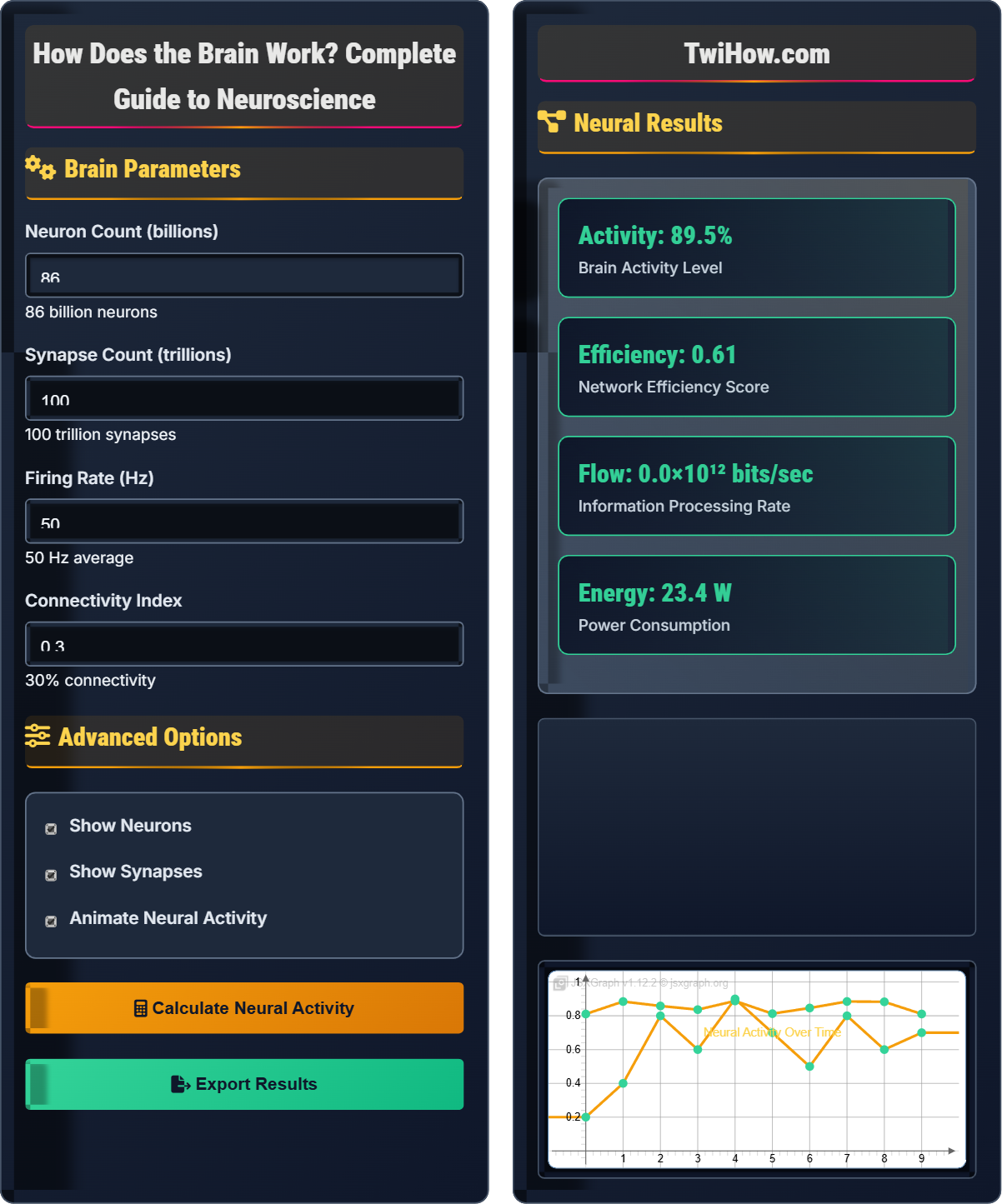
Brain Parameters
Advanced Options
Neural Results
| Region | Function | Neurons | Activity |
|---|---|---|---|
| Cerebral Cortex | Thinking, Consciousness | 16 billion | High |
| Cerebellum | Motor Control | 50 billion | High |
| Hippocampus | Memory Formation | 4 million | Variable |
Where Vm is membrane potential, Vrest is resting potential, Veq is equilibrium potential, and τ is time constant.
How the Brain Works
The brain is the central organ of the nervous system that controls most bodily functions and processes information from internal and external environments. It weighs about 1.4 kg and contains approximately 86 billion neurons interconnected by trillions of synapses. The brain processes sensory information, controls motor functions, stores memories, and enables consciousness and thought.
Where:
- I: Ionic current across membrane
- V: Voltage difference across membrane
- R: Membrane resistance
Additionally, the Nernst equation describes equilibrium potential: E = (RT/zF) ln([ion]out/[ion]in)
Neurotransmitters are chemical messengers that transmit signals across synapses:
- Glutamate: Primary excitatory neurotransmitter
- GABA: Primary inhibitory neurotransmitter
- Dopamine: Reward and motivation pathways
- Serotonin: Mood regulation
- Acetylcholine: Muscle activation and memory
These chemicals bind to specific receptors, opening ion channels and altering the postsynaptic neuron's electrical state.
- Medical Imaging: fMRI, PET, EEG for brain activity
- Therapeutics: Drugs targeting neurotransmitter systems
- Brain-Computer Interfaces: Neural prosthetics
- Psychology: Understanding behavior and cognition
- Artificial Intelligence: Neural networks inspired by brain
Neural Fundamentals
Neurons, synapses, neurotransmitters, action potentials, neural networks, plasticity.
Vm = Vrest + (Veq - Vrest)(1 - e-t/τ)
Where Vm = membrane potential, Vrest = resting potential, Veq = equilibrium potential, τ = time constant.
- All-or-nothing principle for action potentials
- Synapses can be excitatory or inhibitory
- Neural networks exhibit plasticity
- Integration occurs at axon hillock
Real-World Applications
Neurological treatments, psychiatric medications, brain-computer interfaces, cognitive therapy.
- Electroencephalography (EEG) for electrical activity
- Functional MRI for blood flow changes
- Single-unit recording for individual neurons
- Optogenetics for targeted neural manipulation
- Brain activity varies by state (sleep/wake)
- Individual differences affect neural patterns
- Network effects emerge from cellular activity
- Plasticity allows adaptation to injury
Neuroscience Quiz
Which of the following best describes the all-or-nothing principle of action potentials?
The all-or-nothing principle states that once the membrane potential reaches the threshold (typically around -55mV), an action potential will occur with the same amplitude regardless of the strength of the stimulus. If the threshold is not reached, no action potential occurs. This principle ensures reliable signal transmission along axons.
The action potential always depolarizes to about +30mV, then repolarizes back to resting potential, maintaining consistent amplitude and duration.
The answer is B) Action potentials are always the same size once threshold is reached.
This principle is fundamental to neural signaling. It prevents signal degradation over long distances and ensures reliable communication. The strength of a stimulus is encoded in the frequency of action potentials, not their amplitude. This digital-like coding system makes neural communication robust and error-resistant.
Action Potential: Rapid electrical signal along neuron axon
Threshold: Critical membrane potential for AP initiation
All-or-Nothing: Principle of binary signal transmission
• AP amplitude is constant once initiated
• Threshold must be reached for AP
• Signal strength encoded in frequency
• Think of AP like a light switch (on/off)
• Brightness (frequency) varies, not signal size
• Consistent signal prevents degradation
• Confusing amplitude with frequency coding
• Thinking APs can be partial
• Forgetting threshold requirement
Describe the process of synaptic transmission from presynaptic neuron to postsynaptic neuron. Include the role of calcium ions, neurotransmitters, and receptor binding in your explanation.
Synaptic transmission occurs in several steps:
1. Action Potential Arrival: AP reaches presynaptic terminal, depolarizing the membrane.
2. Calcium Influx: Depolarization opens voltage-gated Ca²⁺ channels, allowing calcium to rush into the terminal.
3. Vesicle Fusion: Calcium triggers fusion of neurotransmitter-containing vesicles with presynaptic membrane.
4. Neurotransmitter Release: NTs are released into synaptic cleft via exocytosis.
5. Diffusion: NTs diffuse across synaptic cleft to postsynaptic membrane.
6. Receptor Binding: NTs bind to specific receptors on postsynaptic membrane.
7. Ion Channel Opening: Receptor binding opens ion channels, changing postsynaptic membrane potential.
8. Signal Integration: Postsynaptic changes contribute to overall neuronal activity.
9. Termination: NTs are removed by reuptake, enzymatic degradation, or diffusion.
This electrochemical process bridges the gap between neurons, allowing them to communicate. The calcium-dependent release mechanism ensures that neural signals are transmitted reliably only when an action potential arrives. The diversity of neurotransmitters and receptors allows for complex modulation of neural signals, enabling everything from simple reflexes to complex thoughts.
Synapse: Junction between two neurons
Presynaptic: Neuron sending the signal
Postsynaptic: Neuron receiving the signal
• Calcium triggers NT release
• NTs bind specific receptors
• Signal terminates quickly
• Think of synapse as a relay baton handoff
• Calcium is the "trigger" for release
• NTs are "keys" to receptor "locks"
• Forgetting calcium's role in release
• Confusing presynaptic with postsynaptic
• Thinking transmission is continuous
A stroke patient has damage to Broca's area in the left hemisphere. Explain how neural plasticity might help recovery of speech functions. Discuss the mechanisms of plasticity and factors that influence recovery potential.
Neural plasticity offers hope for stroke recovery through several mechanisms:
Structural Plasticity:
- New synaptic connections can form around damaged areas
- Undamaged neurons can take on new functions
- Existing pathways can strengthen to compensate
Functional Plasticity:
- Right hemisphere may assume some language functions
- Adjacent brain regions can reorganize to support speech
- New neural circuits can develop alternative pathways
Recovery Factors:
- Age: Younger brains show greater plasticity
- Severity: Smaller lesions allow better recovery
- Timing: Early intervention maximizes plasticity
- Practice: Intensive therapy promotes rewiring
- Environment: Stimulating environments enhance plasticity
Recovery involves the formation of new neural pathways that bypass damaged tissue, allowing alternative routes for speech processing to develop.
Neural plasticity demonstrates the brain's remarkable ability to adapt and reorganize. This capacity for change underlies learning, memory, and recovery from injury. The brain's plasticity is greatest in childhood but continues throughout life, though it diminishes with age. Therapeutic interventions can harness plasticity to promote recovery.
Neural Plasticity: Brain's ability to reorganize and form new connections
Broca's Area: Region responsible for speech production
Compensatory Rewiring: Alternative pathway formation
• Plasticity enables recovery from brain injury
• Recovery requires stimulation and practice
• Plasticity decreases with age
• Brain can rewire itself after injury
• Therapy promotes beneficial plasticity
• Repetition strengthens new pathways
• Thinking brain damage is always permanent
• Underestimating plasticity potential
• Forgetting role of rehabilitation
Explain how imbalances in dopamine and serotonin contribute to Parkinson's disease and depression, respectively. Describe the therapeutic approaches for each condition and explain why these treatments work at the molecular level.
Parkinson's Disease (Dopamine Imbalance):
- Caused by death of dopamine-producing neurons in substantia nigra
- Leads to motor symptoms: tremor, rigidity, bradykinesia
- Therapy: L-DOPA (precursor to dopamine) or dopamine agonists
- Molecular basis: L-DOPA crosses blood-brain barrier and converts to dopamine
Depression (Serotonin Imbalance):
- Associated with reduced serotonin activity in certain brain regions
- Leads to mood symptoms: sadness, anhedonia, sleep disruption
- Therapy: SSRIs (Selective Serotonin Reuptake Inhibitors)
- Molecular basis: SSRIs block serotonin reuptake, increasing synaptic levels
Both conditions demonstrate how neurotransmitter imbalances can cause specific symptoms. Treatment aims to restore normal neurotransmitter levels or sensitivity, showing the direct link between chemistry and behavior.
These examples illustrate the biochemical basis of neurological and psychiatric disorders. They demonstrate how specific neurotransmitter systems control particular functions and how pharmacological interventions can target these systems. The success of these treatments validates the chemical imbalance theory of certain conditions.
Neurotransmitter Imbalance: Abnormal levels of chemical messengers
SSRI: Selective Serotonin Reuptake Inhibitor
L-DOPA: Dopamine precursor medication
• Specific NTs control specific functions
• Imbalances cause characteristic symptoms
• Targeted therapy can restore balance
• Different NTs have different functions
• Too much or too little causes problems
• Medications target specific systems
• Thinking all mental disorders are identical
• Confusing different neurotransmitter systems
• Forgetting molecular mechanisms of drugs
Which brain region is primarily responsible for processing visual information?
The visual cortex, located in the occipital lobe at the back of the brain, is the primary region for processing visual information. It receives input from the retina via the optic nerves and processes this information to create our visual perceptions.
The visual cortex is organized retinotopically, meaning adjacent neurons respond to adjacent parts of the visual field. It processes different aspects of vision including color, motion, shape, and depth through specialized subregions.
Other options: Auditory cortex processes sound, motor cortex controls movement, and prefrontal cortex handles executive functions.
The answer is B) Visual cortex.
This demonstrates the principle of functional specialization in the brain. Different regions are anatomically and physiologically optimized for specific functions. The visual cortex's location at the back of the brain is somewhat counterintuitive but makes sense evolutionarily as it's protected by the skull and positioned to receive direct input from the eyes via the optic pathways.
Visual Cortex: Brain region processing visual information
Occipital Lobe: Brain region containing visual cortex
Retinotopic Organization: Mapping of visual field to cortex
• Brain regions have specialized functions
• Location often relates to function
• Sensory areas process specific modalities
• Name often indicates function (visual cortex)
• Sensory areas receive specific inputs
• Motor areas control specific outputs
• Confusing different cortical areas
• Forgetting specific functions
• Misremembering anatomical locations
FAQ
Q: How does the brain store memories?
A: Memories are stored through changes in the strength and structure of synaptic connections between neurons. When we learn something, specific neural circuits are activated repeatedly, strengthening the connections between those neurons. This process, called long-term potentiation (LTP), involves structural changes at synapses including increased neurotransmitter release and enhanced receptor sensitivity. Memories are distributed across multiple brain regions, with the hippocampus playing a crucial role in consolidating short-term memories into long-term storage. Different types of memories (episodic, semantic, procedural) involve different neural networks and molecular mechanisms.
Q: What is the difference between the brain and the mind?
A: The brain is the physical organ composed of neurons, glia, and other cells, while the mind refers to the collection of cognitive functions that emerge from brain activity. The brain is the hardware, and the mind encompasses the software (thoughts, emotions, consciousness, perception). However, this distinction is somewhat artificial since mental processes arise directly from neural activity. Consciousness, often considered the essence of the mind, remains one of neuroscience's greatest mysteries. While we can observe brain activity, the subjective experience of consciousness (qualia) is difficult to explain purely through neural mechanisms. This is known as the "hard problem" of consciousness.
Q: How does the brain process language?
A: Language processing involves several specialized brain regions, primarily in the left hemisphere for most people. Wernicke's area (in the temporal lobe) is responsible for language comprehension, while Broca's area (in the frontal lobe) controls speech production. The arcuate fasciculus connects these regions, allowing for repetition and naming. Written language involves the visual cortex for reading and angular gyrus for converting visual words to sounds. Damage to these areas causes specific language deficits: Wernicke's aphasia (fluent but meaningless speech) and Broca's aphasia (non-fluent but meaningful speech). The brain's language network is highly lateralized and develops through critical periods in childhood.